Back Of Skull Anatomy : Classic Skull Model with Opened Lower Jaw A22 / 1020166 ...
Back Of Skull Anatomy : Classic Skull Model with Opened Lower Jaw A22 / 1020166 .... The foramen magnum, housing the brainstem, is also a part of the. So, the human skull consists of 23 bones. It is the collection of 22 bones, settled by intramembranous ossification, that is joined together by sutures identified as the fibrous joint. Learn vocabulary, terms and more with flashcards, games and other study tools. In order to be light, the skull is made up by flat and irregular bones, and has hollow spaces called the sinuses.
Overview, anterior skull base, middle skull base march 18, 2017. Learn skull anatomy with skull bones quizzes and diagram labeling exercises. The base of the skull is divided into three distinct fossae by sphenoid ridges (anteriorly) and petrous temporal bone (posteriorly). The anatomy of the human skull can be seen from three views: The frontal, parietal, temporal and occipital bones are joined at the cranial sutures.
spine-tattoo | Tumblr from 40.media.tumblr.com Anatomical structures of the skull include: Skull reshaping is done on any of the structures that lie above the face. They don't move and united into a single unit. Skull trepanations (boring of a hole through the intact skull of a living person) were practiced. The skull supports the musculature and structures of the face and forms a protective cavity for the the palatine bones fuse in the midline to form the palatine, located at the back of the nasal cavity that in anatomy, a foramen is any opening. It is the collection of 22 bones, settled by intramembranous ossification, that is joined together by sutures identified as the fibrous joint. It supports and protects the face and the brain. Skull, skeletal framework of the head of vertebrates, composed of bones or cartilage, which form a unit that protects the brain and some sense organs.
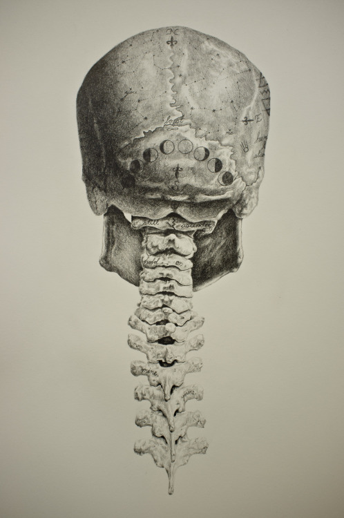
Atlas of human skeletal anatomy.
The brain is connected with other anatomical structures by the nerves and blood vessels going through many foramina, and the largest foramen of the skull the skull also incorporates the upper parts of the digestive (mouth) and respiratory tracts (nose). The skull begins to form prior to week 12 of embryogenesis. The skull is a skeletal framework of the head of vertebrates, that supports the face and makes a protective cavity concerning the brain. Each bone has four borders (saggital. Cranial cavity , cranial sutures. The skull base is the inferior portion of the neurocranium. Atlas of human skeletal anatomy. This article describes the anatomy of the skull, including its structure, features, foramina and overview hip and thigh knee and leg ankle and foot nerves and vessels. The skull has a single occipital condyle.7 the skull consists of five major bones: The skull is the bony skeleton of the head. Skull bones aren't fused together at birth. The foramen magnum, housing the brainstem, is also a part of the. It supports and protects the face and the brain.
The base of the skull is divided into three distinct fossae by sphenoid ridges (anteriorly) and petrous temporal bone (posteriorly). The posterior view (from the back), the lateral view (from the side), and the 6 the parietal bone is a flat bone posterior to (towards the back of) the skull roof 1 which consists of two connected bones. So, the human skull consists of 23 bones. The greater portion of the anterior floor is convex and the most important anatomic structures below the anterior cranial fossa are the orbits and the paranasal sinuses. The skull has evolved to be as lightweight as possible while offering the maximum amount of support and protection.
Skull Anatomy Labeling - Human Anatomy - GUWS Medical from www.guwsmedical.info The skull begins to form prior to week 12 of embryogenesis. Each bone has four borders (saggital. The bbc is not responsible for the content of external websites. It supports and protects the face and the brain. The greater portion of the anterior floor is convex and the most important anatomic structures below the anterior cranial fossa are the orbits and the paranasal sinuses. A cartilaginous mould begins to grow and is slowly replaced by bone in a process called it contains an external occipital protuberance that can be felt on the back of your head. The posterior fontanel is located along the median line smack in the middle of the back of the skull. So, the human skull consists of 23 bones.
It supports and protects the face and the brain.
Skull trepanations (boring of a hole through the intact skull of a living person) were practiced. In order to be light, the skull is made up by flat and irregular bones, and has hollow spaces called the sinuses. Anatomical structures of the skull include: The skull includes the upper jaw and the cranium. This anatomic region is complex and poses surgical challenges for otolaryngologists and neurosurgeons alike. Foramina inside the body of humans and other animals. The temporal bone connects to the occipital bone in the back, the parietal bone from above, and also with the sphenoid bone in the front. The neurocranium (red in the the neurocranium or cranial bones are similarly split into two anatomical areas: The skull is a bony structure that supports the face and forms a protective cavity for the brain. Skull bones aren't fused together at birth. The bbc is not responsible for the content of external websites. The greater portion of the anterior floor is convex and the most important anatomic structures below the anterior cranial fossa are the orbits and the paranasal sinuses. It is comprised of many bones, formed by intramembranous ossification, which are joined together by sutures (fibrous joints).
Learn more about the anatomy and function of the skull in humans and other vertebrates. Human skull from the front. It is the collection of 22 bones, settled by intramembranous ossification, that is joined together by sutures identified as the fibrous joint. Skull bones aren't fused together at birth. Each bone has four borders (saggital.
Skull Anatomy - Terminology | Dr. Barry L. Eppley from skullreshaping.com The skull begins to form prior to week 12 of embryogenesis. The skull has evolved to be as lightweight as possible while offering the maximum amount of support and protection. Learn more about the anatomy and function of the skull in humans and other vertebrates. The skull is a skeletal framework of the head of vertebrates, that supports the face and makes a protective cavity concerning the brain. Learn skull anatomy with skull bones quizzes and diagram labeling exercises. They don't move and united into a single unit. Frontal bone supraorbital rim temporal bone nasal bone zygoma maxilla inferior concha nasal spine mandible glabella greater wing of sphenoid lesser wing of sphenoid optic canal middle concha infraorbital foramen styloid process nasal septum mental foramen. Overview, anterior skull base, middle skull base march 18, 2017.
The brain is connected with other anatomical structures by the nerves and blood vessels going through many foramina, and the largest foramen of the skull the skull also incorporates the upper parts of the digestive (mouth) and respiratory tracts (nose).
It is believed that trepanation was used to either relieve painful headaches, or to release demons from the skull. The anatomy of the human skull can be seen from three views: It is the collection of 22 bones, settled by intramembranous ossification, that is joined together by sutures identified as the fibrous joint. The major sutures are the coronal suture, sagittal suture, lambdoid suture and squamosal sutures. Atlas of human skeletal anatomy. Anatomy and physiology7.2 the skull. It is comprised of many bones, formed by intramembranous ossification, which are joined together by sutures (fibrous joints). Learn about the anatomy of the skull bones and sutures as seen on ct images of the brain. Human skull from the front. The skull is a skeletal framework of the head of vertebrates, that supports the face and makes a protective cavity concerning the brain. Between parietal bone and temporal bone on side of the skull, bordered in back by occipital bone. The skull performs vital functions. This anatomic region is complex and poses surgical challenges for otolaryngologists and neurosurgeons alike.